
 简体中文
简体中文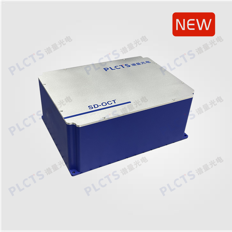
扫一扫
关注公众号
OGS Series SD-OCT Spectrometer delivers 400–1700 nm high-sensitivity, high-throughput spectral detection with 100–200 kHz line rates and OEM customization options
Optical Coherence Tomography (OCT) is a powerful, non-destructive 3D imaging technology widely used in medical diagnostics, biological research, and industrial inspection. With features like non-invasiveness, high resolution, real-time imaging, and depth-resolving capability, OCT systems are typically classified into Time-Domain OCT (TD-OCT) and Frequency-Domain OCT (FD-OCT). The latter includes Spectral-Domain OCT (SD-OCT) and Swept-Source OCT (SS-OCT), both of which offer significant imaging advantages.
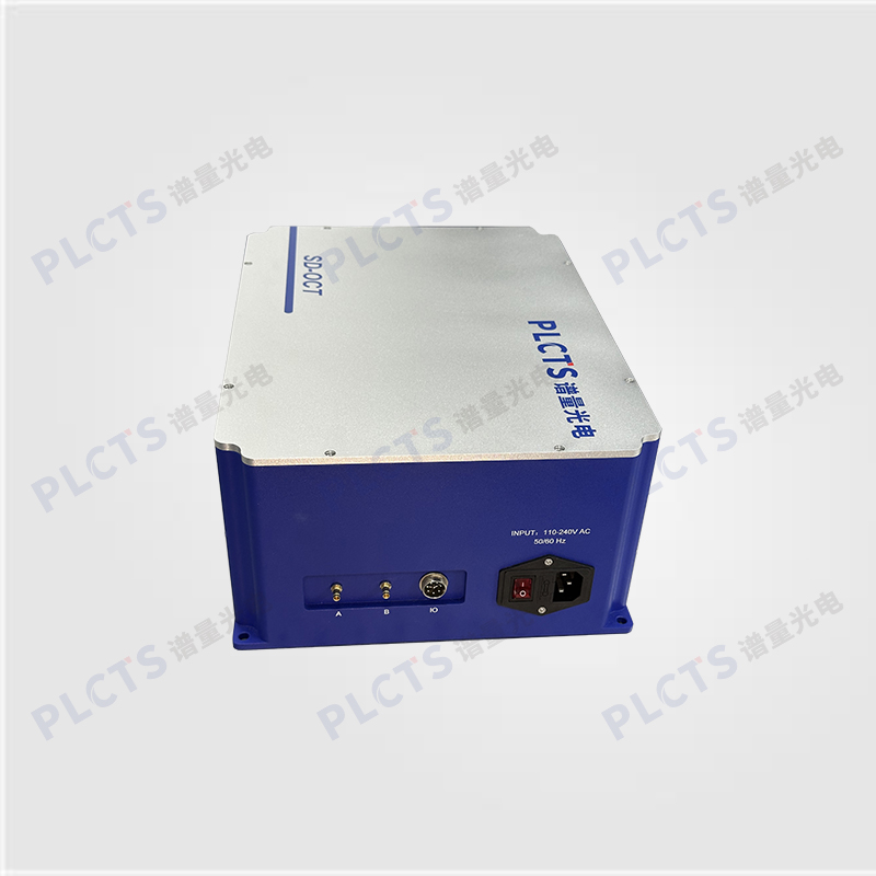
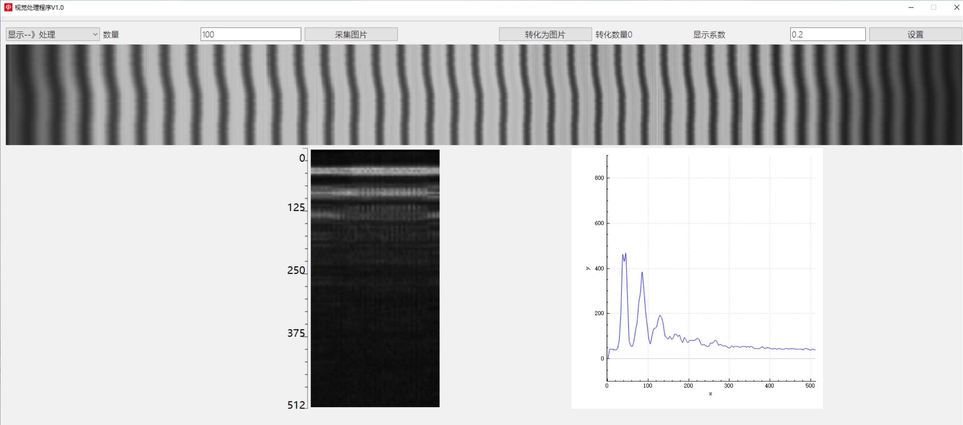
The OGS Series SD-OCT Spectrometer by PLCTS is a high-performance spectral detection system featuring high throughput, high resolution, and high sensitivity. Leveraging a proprietary optical design with VPH gratings and aspheric optics, it minimizes aberrations and delivers superior image quality. Equipped with high-speed cameras covering the visible to near-infrared spectrum (400–1700 nm), it supports line rates up to 100–200 kHz, making it ideal for applications in biological tissue imaging, materials inspection, chemical analysis, and industrial quality control.
To meet integration needs, we also offer OEM customization options, delivering compact and application-specific SD-OCT spectrometers.
With deep expertise in OCT system design, our team has engineered a proprietary high-throughput architecture that overcomes the traditional trade-off between imaging depth and axial resolution. This enables exceptional data quality for deep structures and large-angle scattering, making it a superior choice for basic research, drug testing, and imaging in ophthalmology, dermatology, and angiography.
PLCTS offers customized OCT system solutions,tailored to your application needs. For more information, please contact us.
Note: This product supports evaluation useand can be provided with necessary technical support upon request.
● VPH gratings for low polarization dependence and high diffraction efficiency
● High spatial resolution, enabling sub-micron-level imaging
● Parallel data acquisition for ultra-fast scan speeds
● Non-invasive and real-time imaging capability
● High depth resolution
● Compact design with excellent integration compatibility
● Proprietary algorithms and software for simplified data acquisition and analysis
● Integrated hardware–software system for streamlined operation
● Ophthalmic Imaging:Widely used for high-resolution imaging of the retina, cornea, macula, and related eye structures
● Dermatological Imaging:Detailed structural imaging of skin tissues
● Industrial Inspection:Non-destructive testing of internal microstructures in materials, such as semiconductors and optical fibers
● Medical Research:Applied in tissue studies, disease diagnostics, and cellular structure imaging
● Biological Tissue Research:Including cell morphology, tissue engineering, and drug delivery
● Fundamental Research:Such as photonic interaction studies and surface/interface chemical analysis
|
|
|
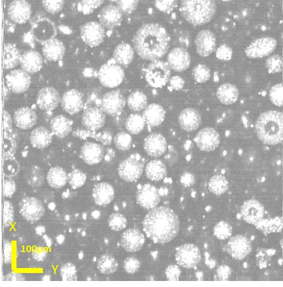
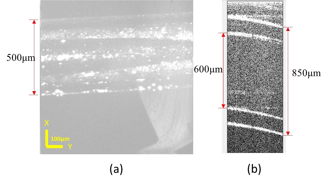
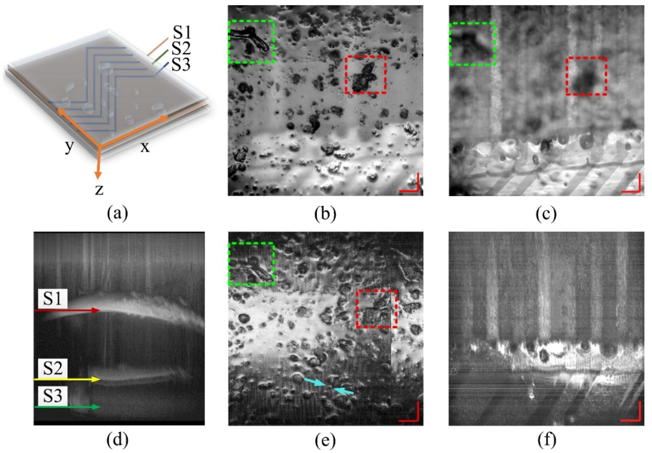
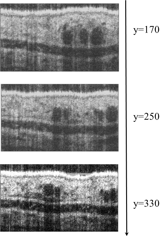
| ↑En face image of a 100μm diameter glass microsphere | ↑Planar and cross-sectional views of a fluidic pipeline. | ↑Laminated layer tomography image of an LCD panel. |
|
|
|
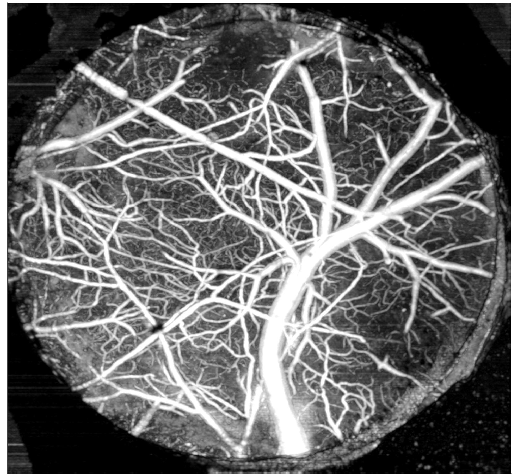
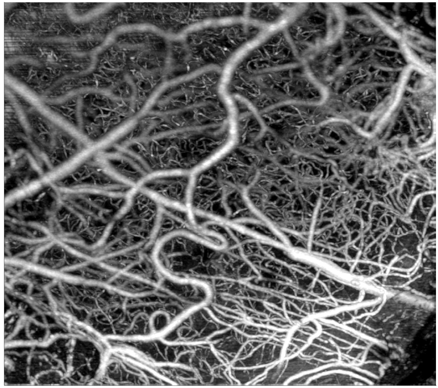
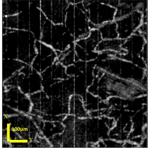
| ↑Cross-section of a mouse ear | ↑Mouse brain angiography imaging1 | ↑Mouse brain angiography imaging2 |
↑Angiography image of a mouse ear.1
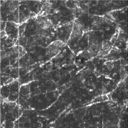
↑Angiography image of a mouse ear.2
PL Optics offers customized OCT system solutions tailored to your specific application needs. For more details, please contact us.
Note: This product is available for trial and necessary technical support will be provided!