
 简体中文
简体中文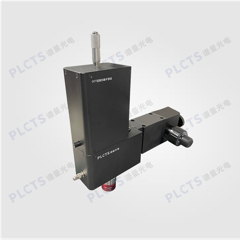
扫一扫
关注公众号
Compact Michelson scanning system with up to 200 kHz A-scan, 5 mm depth, 400–1700 nm spectral range.
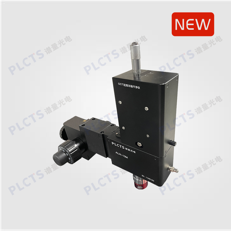
PLCTS SD-OCT Michelson Interferometric Scanning System is a compact and integrated solution designed for spectral-domain OCT applications. It features a complete internal interferometric setup—including a movable reference arm, sample arm, fast/slow axis galvanometers, and a microscope probe. The broadband OCT light source is fiber-coupled into the system and works seamlessly with our OGS series spectrometers to enable high-resolution 2D cross-sectional and 3D volumetric imaging.
Widely used in medical diagnostics, biological research, and industrial inspection, PLCTS provides turnkey and customizable OCT solutions, from full systems to core modules.
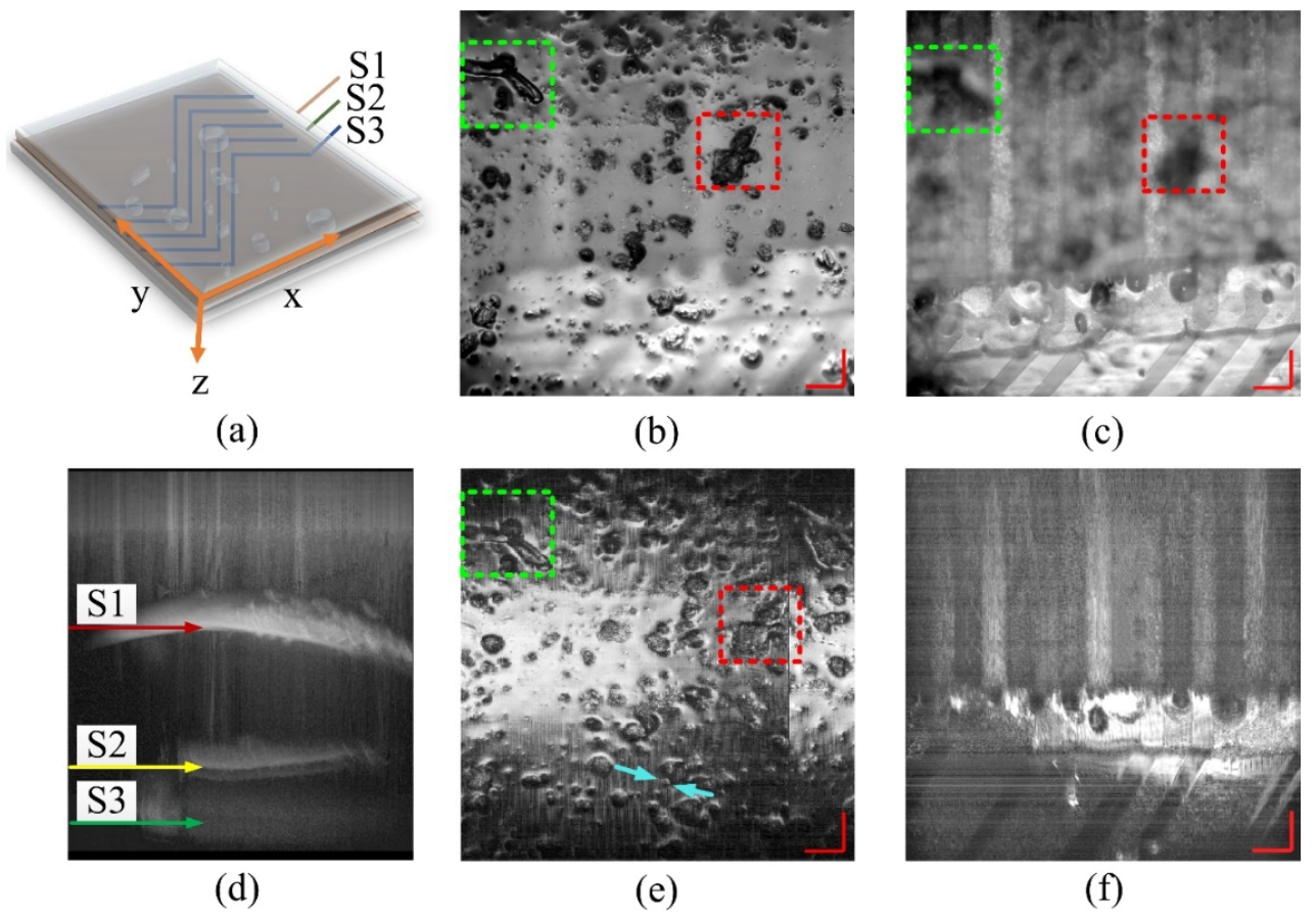
The reference arm is enclosed near the sample to ensure stable phase alignment with the sample arm. Both optical path length and intensity of the reference arm are adjustable to accommodate various imaging conditions (e.g., underwater imaging). To minimize image distortion from dispersion, the reference and sample arms are precisely matched in optical length, and our patented dispersion compensation algorithm effectively corrects sample-induced dispersion (e.g., from water or glass), significantly enhancing imaging resolution.
● Beam-splitting module
● 2D lateral galvanometer scanning system
● Microscope probe
● Reference arm, sample arm, and combiner
● Optical path length adjustment module for reference arm
● Medical diagnostics
● Biological and life sciences
● Industrial non-destructive testing
● Material and structural analysis
● OCT system development and integration
| Model | Supported Wavelength Range | Adjustable Reference Arm | Fiber Interface | Objective |
|---|---|---|---|---|
| STG | 550nm/840nm/1060nm/1310nm/1550nm | 10 mm Travel Range, 2 μm Resolution | FC/APC | 20× |
|
|
|
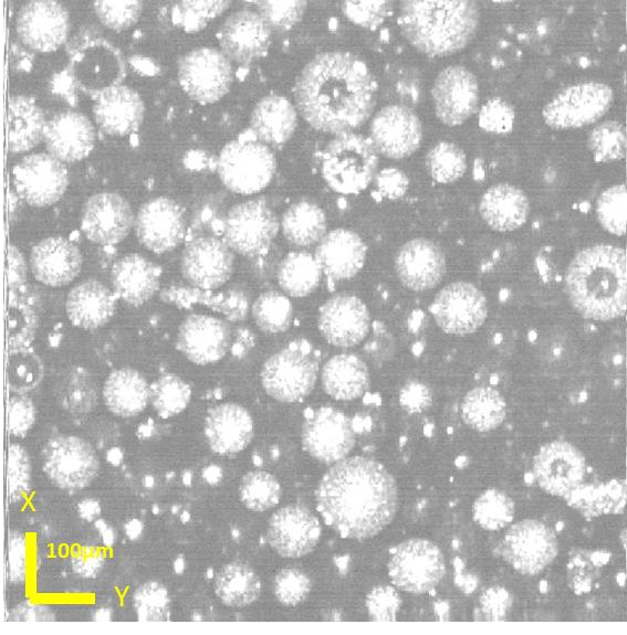
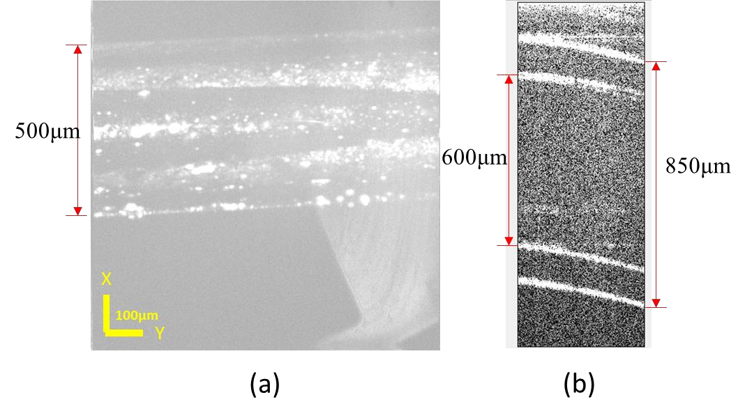
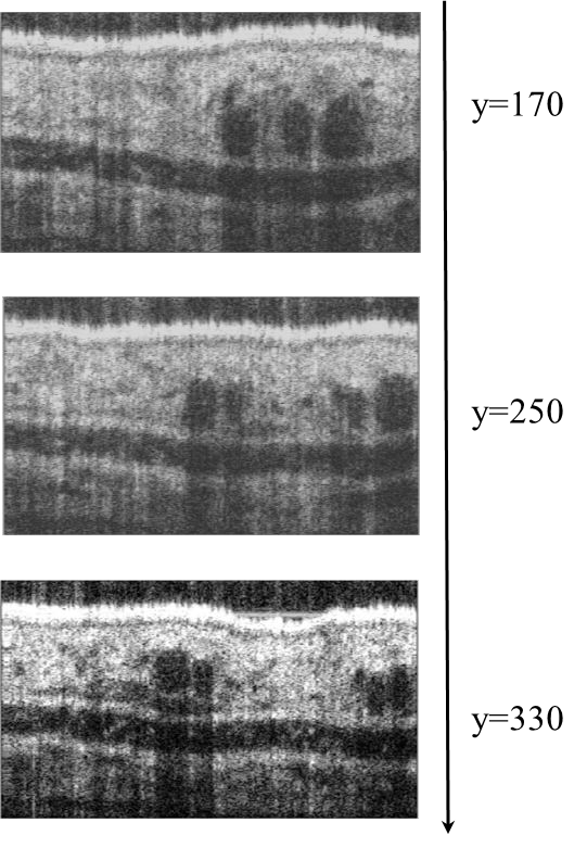
| ↑En face image of a 100μm diameter glass microsphere | ↑Planar and cross-sectional views of a fluidic pipeline. | ↑Laminated layer tomography image of an LCD panel. |
|
|
|
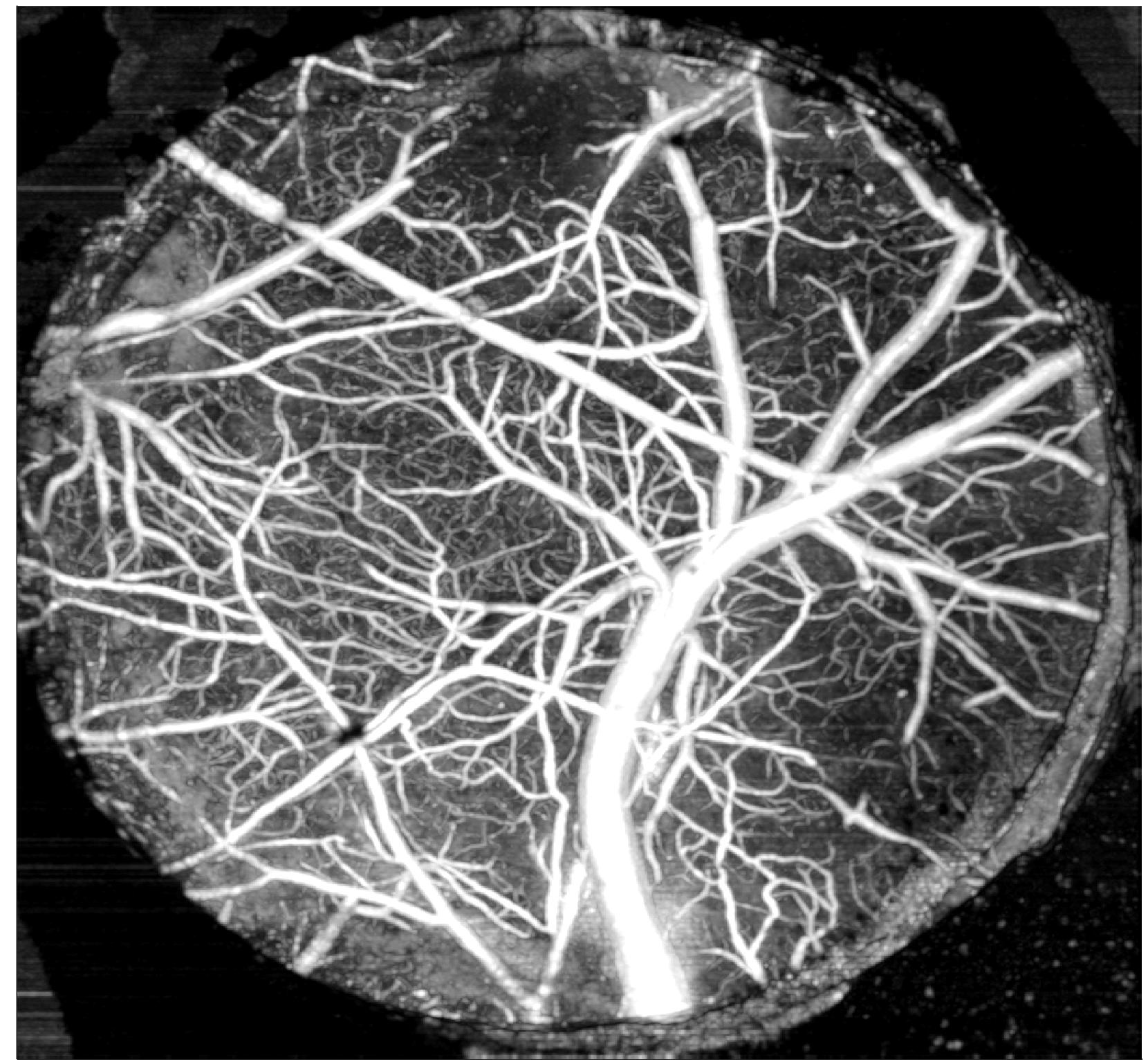
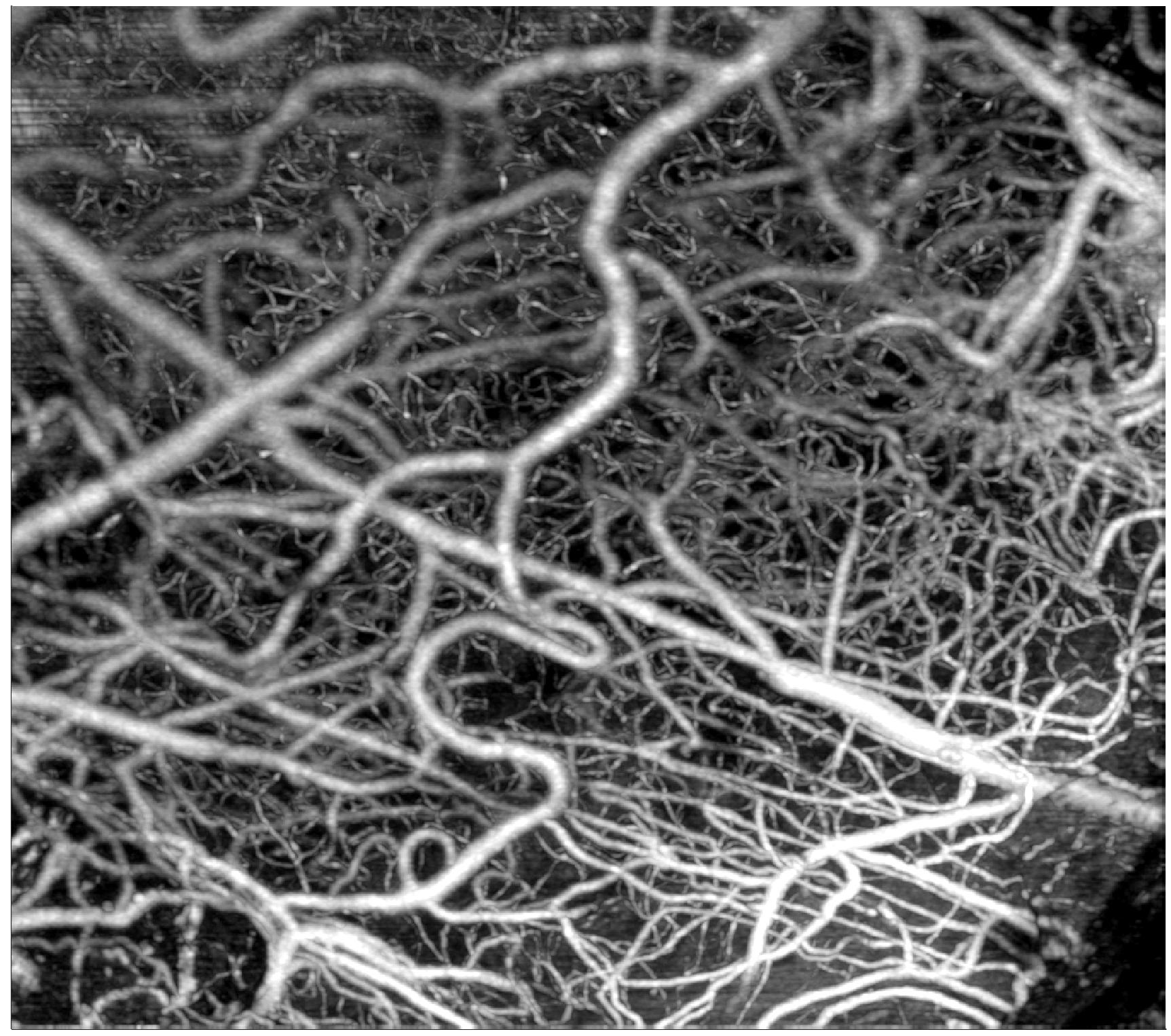
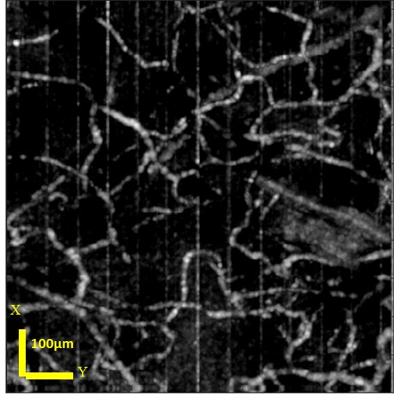
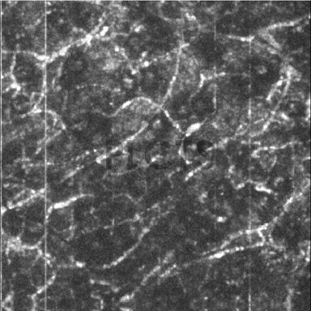
| ↑Cross-section of a mouse ear | ↑Mouse brain angiography imaging1 | ↑Mouse brain angiography imaging2 |
↑Angiography image of a mouse ear.1
↑Angiography image of a mouse ear.2
PL Optics offers customized OCT system solutions tailored to your specific application needs. For more details, please contact us.
Note: This product is available for trial and necessary technical support will be provided!