
 简体中文



扫一扫
关注公众号
Confocal Micro-Raman Spectroscopy System
The Prms Confocal Micro-Raman Spectroscopy System is a high-end micro-Raman materials characterization platform independently developed by PL Optics, building on years of experience in spectroscopic instrument R&D and industry applications.
The system combines ultra-high-resolution optical microscopy with high-precision Raman spectroscopy and incorporates an integrated, high-stability microscopic measurement optical path together with a high-speed, high-resolution area-scanning core module. It comes standard with a large-travel, high-precision motorized XY scanning stage for micrometer-scale, high-resolution point-by-point Raman Mapping acquisition. A closed-loop motorized Z-axis module is also integrated with our proprietary intelligent vision-based focusing algorithm to automatically compensate for defocus caused by sample topography and optical-path drift. This minimizes invalid acquisitions and spectral signal attenuation or distortion, ensuring uniform and complete spectral data across the entire scan area with stable and highly reproducible results. The system is widely applicable to nondestructive microscale characterization of 2D materials, functional thin films, optoelectronic semiconductors, and related samples.
Technical Advantages:
- Fiber-optic confocal technology for a high signal-to-noise ratio
- Multi-level adjustable excitation power for different sample types
- LED Köhler illumination for a uniform field of view and clear imaging
- 532 nm / 785 nm dual-wavelength optical path configuration
- Integrated motorized Z-axis module with intelligent autofocus
Applications:
- Structural characterization of low-dimensional materials
- Microscale analysis of semiconductor optoelectronic devices
- Micro/nanostructure inspection of optoelectronic devices
- Characterization of microscopic material distribution and defects
- Structural studies of catalytic materials for new-energy applications
System Details:

Technical Specifications:
Typical Test Data:

Silicon Wafer ↑

Carbon Nanotube ↑

Graphene ↑

Graphene ↑
Typical 2D Material Test Results
Monolayer WSe₂
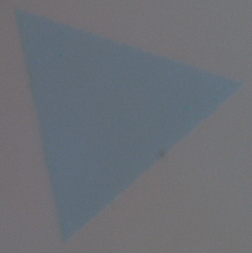
(Monolayer WSe₂ Bright-Field Image)
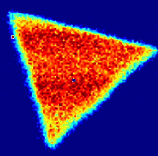
(Monolayer WSe₂ Raman Mapping Image)
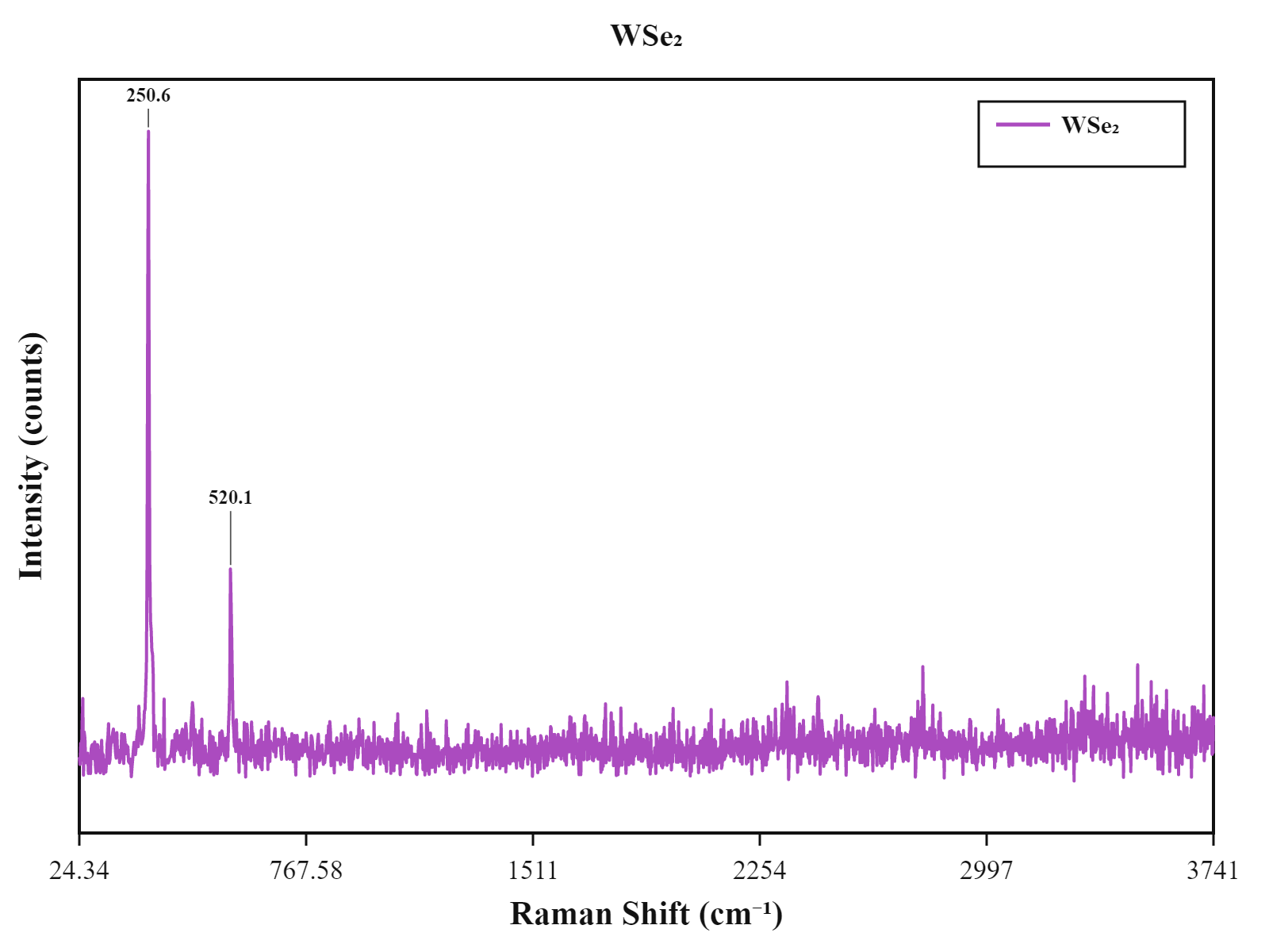
(Monolayer WSe₂ Raman Spectrum)
Monolayer MoS₂
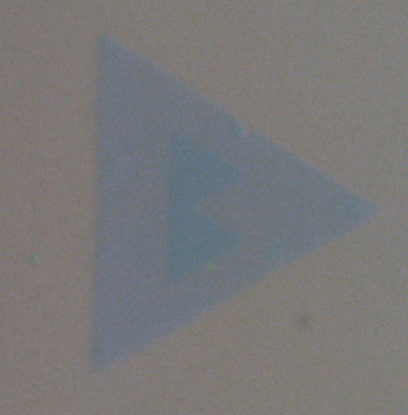
(Monolayer MoS₂ Bright-Field Image)
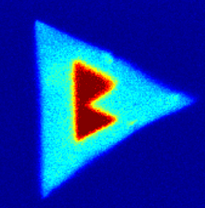
(Monolayer MoS₂ Raman Mapping Image)
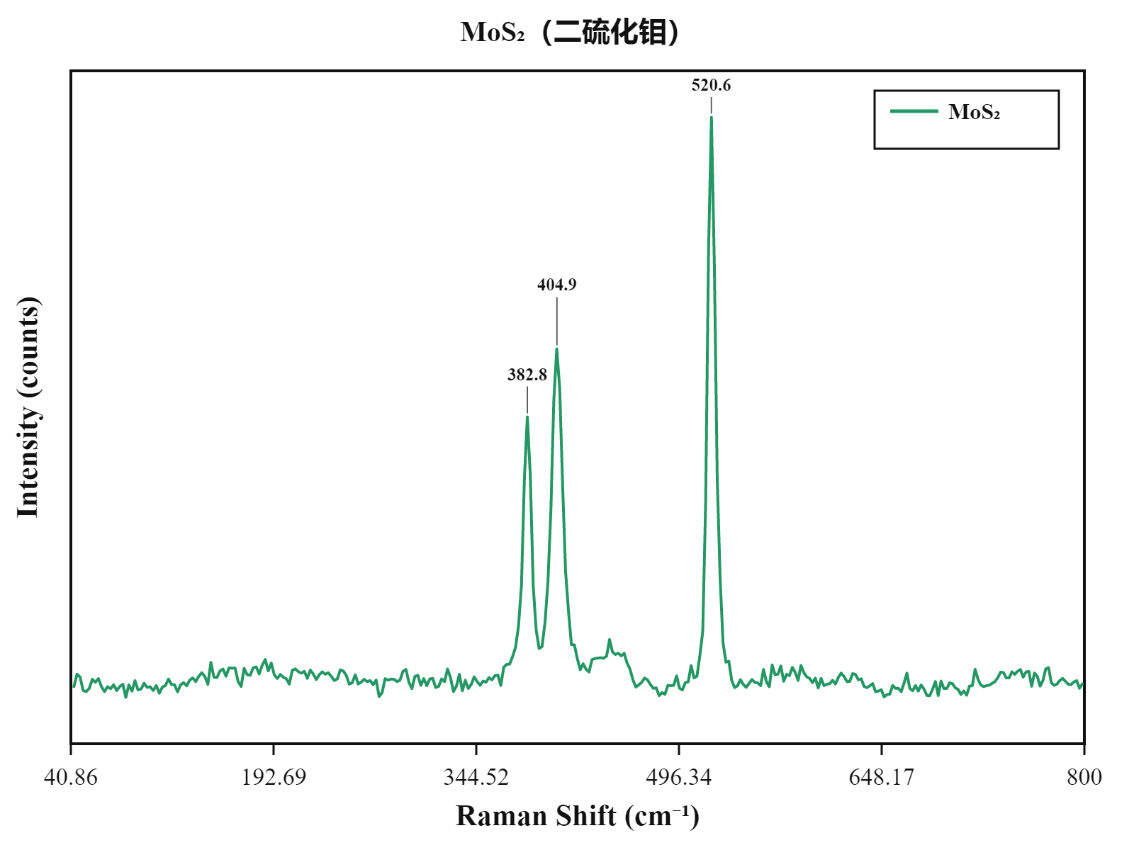
(Monolayer MoS₂ Raman Spectrum)
Monolayer WS₂
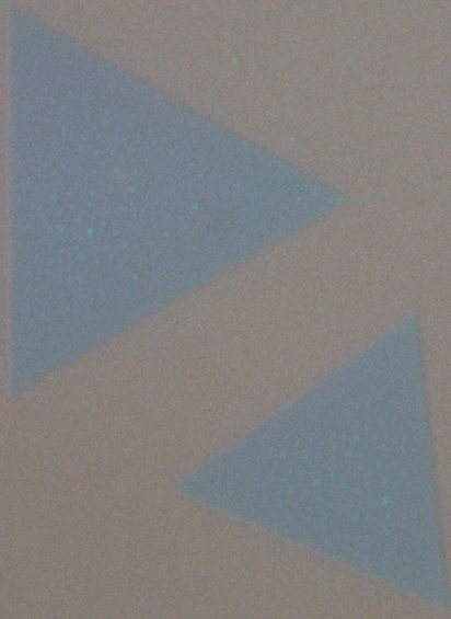
(Monolayer WS₂ Bright-Field Image)
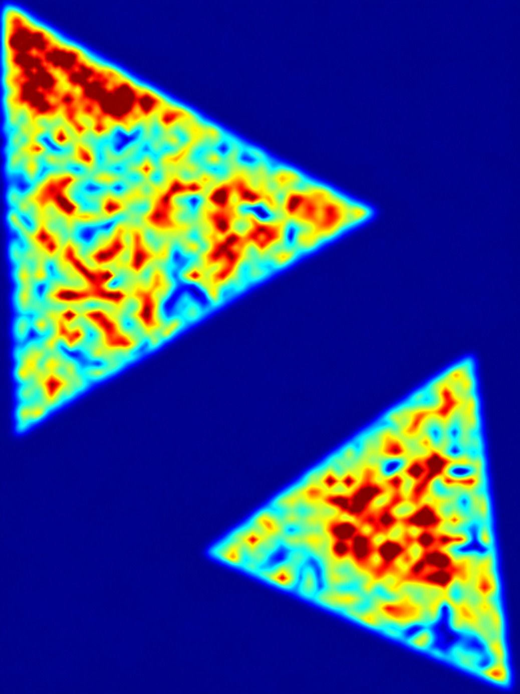
(Monolayer WS₂ Raman Mapping Image)
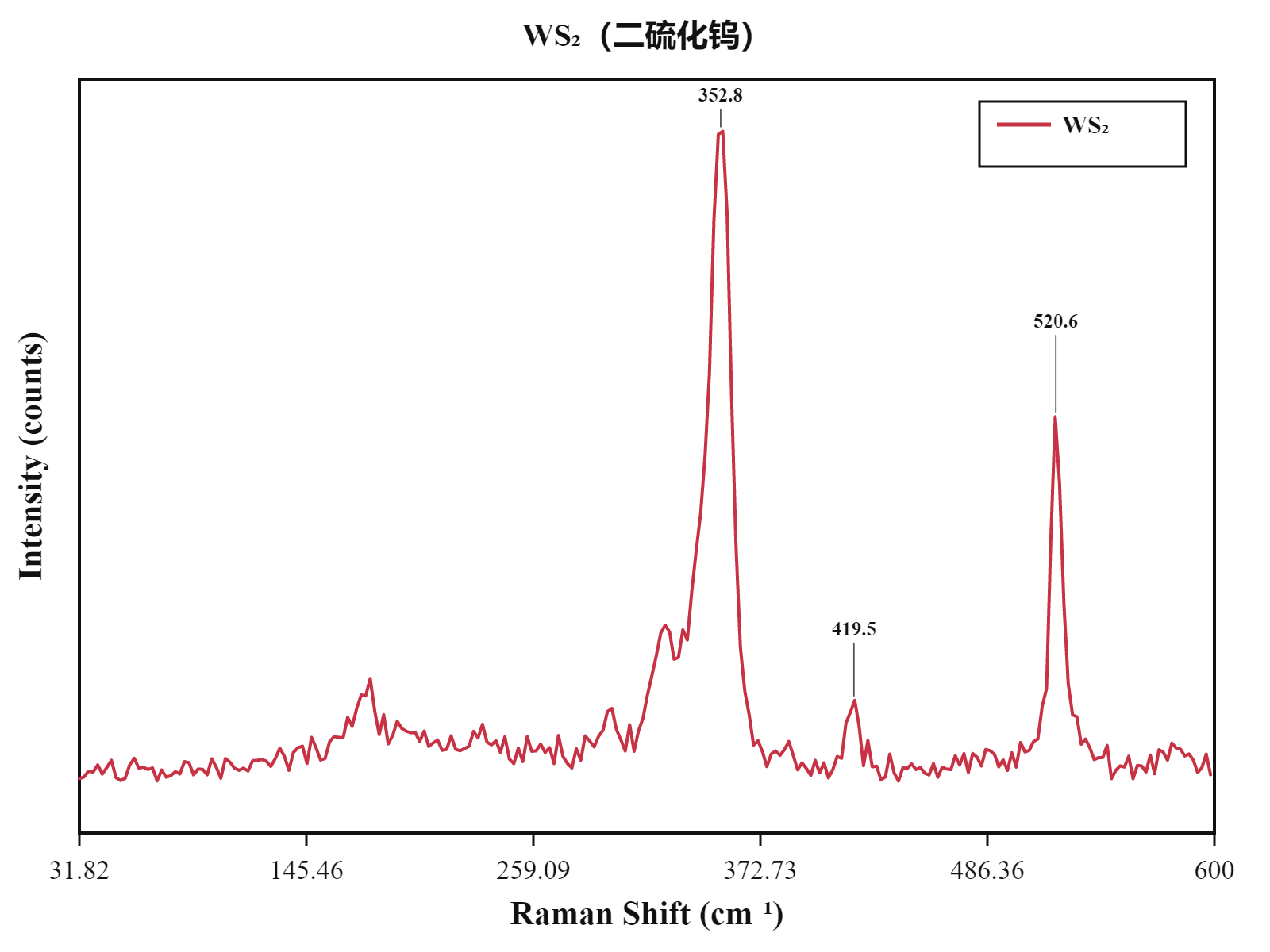
(Monolayer WS₂ Raman Spectrum)
Prms can be customized and upgraded according to application requirements. We also provide a complete range of microscale optical characterization solutions, including micro-Raman spectroscopy, micro-fluorescence spectroscopy, second-harmonic generation (SHG), microscopic photocurrent measurement, and microscopic transmission/reflection spectroscopy. Please contact us for further details.